Diagnostic imaging
Diagnostic Imaging
Magnetic Resonance Imaging (MRI)

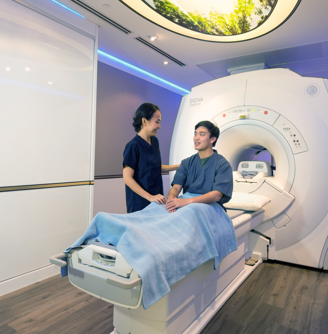
MRI uses waves and a strong magnetic field to produce clear and detailed pictures of the body, without the use of x-rays. It is a non-invasive way of viewing organs, soft tissues, bone and other internal body structures.

  • Analysis of any part of the body including the brain, spine, joints, bones and soft tissues.
  • Diagnosis of small tears, injuries to tendons, ligaments, muscles and small fractures which cannot be seen on x-rays.
  • Diagnosis of degenerative disorders such as arthritis
  • Analysis of damage to the spinal cord after trauma
  • Diagnosis and analysis of infections and tumours of the nervous system, bones and joints.
  • No special preparation of food restriction is required except for MRI Abdomen, MR Liver and MRCP, 4-6 hrs fasting is required before the examination.
  • Inform our staff if you have any existing medical conditions or electronic devices in your body. Please also bring along any information on the implants you have. Surgical screws, staples generally are safe for MRI if they have been placed more than 6 weeks ago.
  • Jewellery or any metal objects must be removed prior to the MRI scan.
  • If you are claustrophobic, sedation may be required. Please highlight this when making an appointment or speak to your doctor about it.
  • A contrast medium may be administered to enhance the visibility of certain tissues or blood vessels. This is usually injected in the vein of your hand or arm.
  • You may be asked to do a blood test for Creatinine Serum before the scan appointment, if required.
  • Please bring along any previous scan images/report (if any)

MAGNETOM Lumina, RadLink's new 3T Open Bore MRI system that delivers productivity and greater patient experience.

Click here to learn more.

Computed Tomography (CT)

A CT (Computed Tomography) scan is performed using an advanced X-ray technique that uses X-ray and computer technology to obtain cross-sectional images of the body, giving detailed information for diagnosis. CT scan images are able to provide more detailed images than plain X-rays. You will be asked to lie down on the CT scan table. During the CT procedure, the CT scan table will move in and out of the scanner several times to capture images of your body.

  • Study of the head and neck, chest, abdomen and pelvis as well as the skeleton.
  • Diagnosis of cancers, such as lung, liver and pancreatic cancers; to confirm or exclude the presence of tumour, evaluate its size and location.
  • Identification of injuries to internal organs in case of trauma.
  • A significant role in the detection and diagnosis of cardio-vascular diseases.
  • 4 hrs fasting is required for CT Enterography and CT Colonography
  • A contrast medium may be administered to highlight relevant areas. Depending on the region to be scanned, you may be given oral contrast or intravenous contrast as ordered by your doctor.
  • If you are on a diabetic medication called Metformin (Glucophage), please do not take it on the day of scan. Resume Metformin (Glucophage) 48 hours after your CT procedure.
  • Inform the radiographer if you suffer from diabetes, mellitus, asthma, heart condition, kidney problems and any drug allergies.
Ultrasound

Ultrasound (Sonography) is an imaging method that uses sound waves to obtaining real-time images within the body. No X-rays or injections are used in ultrasound imaging which makes this modality an extremely safe and painless diagnostic tool.

  • Examination of superficial organs like breast, thyroid and shoulder joints.
  • Analysis of abdominal organs (i.e liver, gallbladder, spleen, pancreas, kidneys, bladder) and pelvic organs (i.e prostate, uterus and ovaries).
  • To evaluate blood flow and assess for blockage or clots.
  • For upper abdomen ultrasound of liver and gallbladder, 4-6 hrs fasting is required.
  • For Pelvis scan (uterus, ovaries, bladder), a full bladder is required to properly visualize the organs. You may have to drink up to 6 glasses of water and avoid emptying the bladder until the examination is complete.
  • Inform the clinic when you make appointment if you are on restricted fluids or suffer from diabetes mellitus.
Mammography


Mammography is a specialised medical imaging of the breasts using a low-dose X-ray system. It is used to aid in the early detection and diagnosis of breast diseases. Together with physical breast examination, it has proven to be the safest and most effective method to screen for early breast cancer.


RadLink also offers 3D mammography (tomosynthesis) a cutting edge technology that increases breast cancer detection; decreases recall rate and aids in identifying size, shape and location of the breast abnormalities.

  • Screening mammography is used to look for possible breast cancer when you are well and have no obvious breast symptoms. It is recommended to do the scan every year for women aged 40 and above and recommended to do the scan every two years for women aged 50 and above.
  • Diagnostic mammography is used when there is a breast symptom such as breast pain, a lump or discharge from the nipple.
  • If you are pre-menopausal, book your appointment during the mid-menstrual cycle when your breasts are usually less tender.
  • Do not wear deodorant, talcum powder or lotion under your arms on the day of examination.
  • Inform the radiographer of any significant past medical history before the start of the examination.
  • Bring your old mammogram films, if any, for comparison.
Bone Mineral Densitometry (BMD)

BMD is the method used to quantify the mass of bone in the body. It is also effective in tracking the effects of treatment for osteoporosis and other conditions that cause bone loss.

BMD is often used to diagnose osteoporosis. This is a common bone disease which makes bone fragile and easy to fracture. Some common causes of osteoporosis include:


  • Lack of physical activity
  • Low calcium intake in the diet
  • Post-menopausal
  • An alcoholic or/and smoker
  • Inform the radiographer if there is any metal in your body, such as spinal fusion rods or a hip prosthesis.
  • It cannot be scheduled within 4 days following any contrast scans such as barium studies, IVU, CT scan or radio-isotope nuclear medicine scans.
  • No special preparations or appointment booking is required for BMD scan.
X-Ray

X-ray is a conventional technique used for the visualization of the bones and soft tissue structures in the body. It uses X-rays to create 2D images of the human body.

  • It is useful to diagnose bone and joint injury and diseases such as fractures, dislocations and infection. The imaging process is painless, fast and uses very low doses of radiation, making X-ray a very quick and reliable diagnostic tool.
  • A chest x-ray is usually done for the evaluation of lungs, heart and surrounding anatomy.
  • No appointment is required for this examination.
  • No special preparation is required
RadLink opening hours
​All RadLink Centres

Monday - Friday: 8:30AM – 5:30PM

Saturday: 8:30AM – 12:30PM

Closed on Sundays and Public Holidays

All RadLink Lite Centres

Monday – Friday: 8:30AM – 5:00PM (Lunch hour: 1:00PM – 2:00PM)

Saturday: 8:30AM – 1:00PM

Sunday: 9:00AM – 1:00PM (Ang Mo Kio only)

Radlink front desk
Drop us a message!

    radlink contact us image